


To date, science knows about 280 species of worms that can develop and live in the human body, parasitizing in various organs and tissues. The frequency of human worm infections depends on the climatic and socio-economic conditions of specific areas (in underdeveloped countries, especially in those located in the tropics and subtropics, the level of parasitic infections is much higher than in economically developed countries).
Ways to infect humans with helminths
- Biohelminthiasis (animal infection).
- Infectious helminthiasis (human-to-human transmission).
- Geohelminthosis (diseases caused by parasites that carry out one of their life cycles in the earth).
Factors influencing the manifestations of helminthiasis
- The way the parasite enters the body;
- The degree of adaptation of the helminth to the human body;
- Population density (number) of parasitic individuals;
- The habitat of the worm (tissue parasites live in the thickness of soft tissues, and luminal - in the lumens of hollow organs). Some helminths in different phases have both luminal and tissue form. The larval and developing stages of worms, as a rule, cause more pronounced pathological changes.
In the absence of re-infection, the number of adult parasites in the human body does not increase. This feature significantly distinguishes helminth infestations from diseases caused by bacteria, viruses, fungi and protozoa.
Human worms: symptoms
Helminthiasis is a disease characterized by 2 stages (acute, from two weeks to two months) and chronic (from several months to several years).
Symptoms of the acute phase of helminthiasis
The first signs of the disease may appear at different times (most often after 2-3 weeks, in ascariasis - after 2-3 days, and in filariasis, the incubation period can last 6-18 months).
In the acute stage of parasitic invasion, the most characteristic symptom is an allergic reaction (antibodies to antigens of migrating larvae of parasites are produced). Often in people infected with worms, itchy skin rashes appear, prone to recurrence, enlarged regional lymph nodes, generalized or local edema, muscle and joint pain may occur. Also, migrating larvae of parasites can cause chest pain, cough, choking attacks, stool disorders, nausea and vomiting.
At the same time, the acute phase of helminthiasis may be accompanied by more serious disorders (severe forms of pneumonia, hepatitis, allergic myocarditis, hepatosplenomegaly (enlarged liver and spleen), meningoencephalitis).
The number of eosinophils in the blood increases (eosinophilia) and the normal ratio between protein fractions is disturbed (dysproteinemia).
Signs of chronic helminthiasis
The symptoms of the chronic phase directly depend on which organ is "inhabited" by parasites, as well as their size and number play an important role.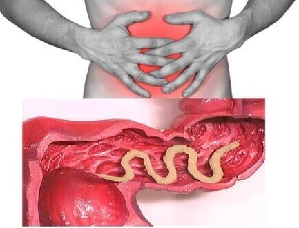 So, when it parasitizes in the intestines of individuals, the disease can be asymptomatic (except in cases of infection with very large parasites). The characteristic signs of the chronic phase of intestinal helminthiasis are dyspeptic disorders. In children, asthenoneurotic and pain syndromes are more pronounced. With massive invasion of roundworms, the development of intestinal obstruction, obstructive jaundice and pancreatitis is possible.
Consuming all the substances necessary for their vital activity from the body of the host, helminths cause digestive disorders, impaired absorption of vitamins, minerals, carbohydrates, proteins and fats. At the same time, the waste products of worms inhibit the normal intestinal microflora and reduce the body's immune system.
In people suffering from helminthiasis, due to a weakened immune system and increased cell division (a consequence of the constant repair of tissues damaged by parasites), the risk of malignant tumors increases significantly.
Types of helminths parasitizing the human body
The causes of human helminthiasis are 2 types of worms: roundworms (nematodes) and flatworms (tapeworms and flukes).
Roundworms
Blade
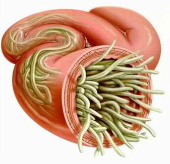
The parasites that cause enterobiosis are small (up to 10 mm) thin hollow worms with a grayish-white coloration. Infection occurs through food (by mouth). The reason for this is dirty hands. The eggs of the parasite can be in the ground, on the wool of infected animals, unwashed vegetables and fruits and more. At the same time, in case of enterobiosis, cases of self-infection are common (especially in children), as a result of scratching the itchy areas and the subsequent ingestion of eggs. 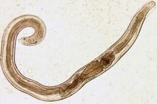 The larva of the squirrel develops within two weeks in the digestive tract. Once an adult, the worm parasitizes the lower parts of the small and upper parts of the colon.
Even in the larval stage, the pinworm begins to damage its host's body by producing enzymes that irritate the intestinal walls and lead to the development of an inflammatory process. Adult parasites adhere to or penetrate into the deeper layers of the intestinal mucosa, disrupting its integrity and contributing to the attachment of secondary bacterial infection. In case of perforation of the spines on the wall of the small intestine, peritonitis may develop. Also, due to irritation of the intestinal receptors, the motor and secretory functions of the gastrointestinal tract are disrupted, which leads to the formation of gastroduodenitis, enteritis and others. In childhood, long-term enterobiosis can cause nervous disorders and physical retardation.
Ascaris
Ascaris is a large spindle-shaped parasite with a reddish-yellow color, reaching 40 cm (female) and 15-25 cm (male) in adulthood. Without suction cups or other fasteners, the roundworm can move on its own to food masses. The eggs laid by the female of the parasite are excreted together with the faeces.
Infection with ascariasis occurs when mature eggs are ingested with water or unwashed vegetables and fruits with soil particles. Once the eggs enter the intestines, mature larvae emerge. Then, penetrating the intestinal wall, they reach the heart through the bloodstream and from there enter the lungs. Through the lung alveoli, the roundworm larva enters the oral cavity again through the airways. After repeated ingestion, the parasite reaches the small intestine, where it develops into an adult. The worm lives for 12 months, after which it dies and is excreted in the faeces. Both one and several hundred individuals can live in the intestines of a host.
In the intestinal phase of their existence, roundworms, endowed with the ability to spiral movements, can penetrate even the narrowest openings. This characteristic of the parasite often leads to the development of quite serious complications (obstructive jaundice or pancreatitis). Allergens secreted by roundworms can provoke severe allergic reactions. A large number of adults can cause intestinal obstruction, and worms that get into the airways sometimes cause suffocation.
Vlasoglav
Vlasoglav, the cause of trichocephaly, is a white helminth that parasitizes in the initial part of the colon and reaches a size of 4-5 cm. The parasite feeds on blood and tissues of the rectal mucosa.
The eggs of the whips laid by the female on the intestinal walls come out with the feces. Their development takes place in the environment (optimally in the soil). Eggs with the mature larvae of the parasite enter the body through food, through dirty hands, with water or unwashed vegetables and fruits.
In a small number of worms, trichocephaly is asymptomatic. In the severe stage (with massive invasion) the patient develops abdominal pain, severe diarrhea develops, sometimes accompanied by rectal prolapse. This condition is most common in exhausted children. In the moderate phase of trichocephaly, growth retardation of the child is possible.
Trichinella
The causative agent of trichinosis is a small round helminth, reaching 2-5 mm in length. Infection occurs when eating poorly roasted meat (pork, bear meat, wild boar).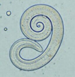 Penetrating into the intestine, the larva of the parasite matures in 3-4 days to the state of a sexually mature individual. The lifespan of the worm is 40 days, after which the parasite dies. By piercing the intestinal wall, the larvae enter the bloodstream and are transmitted to all organs of the human body, settling in the muscles. In this case, the respiratory and facial muscles, as well as the flexor muscles of the limbs are most often affected.
In the first days after the invasion, patients complain of abdominal pain. Then, after about 2 weeks, the body temperature rises to 39-40 C, itchy rashes appear on the skin, muscle pain develops and the face swells. During this period, in case of a massive infection, there is a significant risk of death. After about a month, the patient recovers. The parasite encapsulates in a spiral shape and then dies within two years.
Hookworm and some
These two parasites are similar in biological characteristics as well as in caused diseases. In this regard, it is common to combine under a common name (hookworm). Worms, reaching lengths of 10-15 mm, parasitize in 12-p. intestine. It should be noted that this is one of the most common, but at the same time quite rare parasites. Worm larvae penetrate the human body through the skin in contact with contaminated soil. In addition, entering the bloodstream, they, like roundworms, migrate to the lungs and then through the bronchi, along with sputum expectorant, into the digestive tract. The hookworm parasitizes in the intestine, attaching to the intestinal wall. The parasite, which feeds exclusively on blood, bites through blood vessels that penetrate the mucosa, injecting an anticoagulant component there. The average adult can absorb 0, 05-0, 35 ml of blood per day. Therefore, the most characteristic symptom of this helminthiasis is iron deficiency anemia, as well as a change in the ratio of protein fractions (dysproteinemia).
Flatworms
Wide bar
This is one of the largest helminths, reaching a length of 10-20 meters. The disease caused by this parasite is called diphyllobotriasis. The development cycle of the worm begins with freshwater fish or crustaceans. The larva enters the human body, which is the ultimate owner of the broad tapeworm, along with eggs or infected fish fillets. Reaching the small intestine, the parasite attaches to its wall and grows to a mature individual within 20-25 days.
Diphyllobotriasis occurs against the background of digestive disorders and B12-deficient anemia.
Liver methyl
The parasite that causes opisthorchiasis is a flatworm, reaching a length of 7-20 mm. It should be noted that more than 50% of cases of liver fluke infection (also called feline fluke) occur in Russians. The larvae of the parasite begin to develop after the eggs fall into fresh water (from the snails that have swallowed them). Then they penetrate the body of the fish (carp, crucian carp, bream, roach). Human infection occurs when eating contaminated fish meat that has not undergone sufficient heat treatment. The larva of the hepatic flux from the small intestine penetrates the bile ducts and the gallbladder, and is fixed there with the help of two suction cups.
In the acute phase of helminthiasis, the patient has upper abdominal pain, fever, nausea, muscle aches, diarrhea and skin rashes. The chronic course of opisthorchiasis is manifested by symptoms of hepatitis, inflammation of the bile ducts, cholecystitis, disorders of the digestive tract, nervous disorders, weakness and increased fatigue. The parasite leads to the development of irreversible changes and even after its expulsion the patient does not suffer from chronic inflammatory processes and functional disorders.
Beef and pork tapeworms
These parasites, almost identical in structure, reach a length of 5-6 meters. Infection with teniarinosis and teniasis occurs due to the consumption of beef or pork infected by Finns (one of the intermediate forms of helminthiasis). Viable Finns, presented in the form of whitish bubbles, reaching a size of 0, 5 cm, attach to the wall of the small intestine of man and become an adult in 3 months. The tapeworm, consisting of more than 2, 000 segments, is constantly growing. In this case, the end segments containing the eggs break off and move independently along the colon to the anus and then crawl out of the anus or are released into the external environment along with the stool. The most characteristic symptoms of helminthiasis are disorders of the digestive tract.
Echinococcus
For this parasite, man is an intermediate host. The worm parasitizes on the human body in the form of Finns. The ultimate owner of an echinococcus is a wolf, dog or cat. 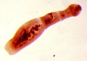 Infection occurs through food through contact with animals and environmental objects seeded with echinococcus eggs. After entering the intestine, oncospheres (six-attached larvae) develop from them. From the intestines they enter the blood and are transmitted to the body.
The worm's "favorite" parasitic sites are the liver and lungs. Sedimenting in these organs, the larva becomes a fine (echinococcal cyst), which gradually increasing in size, begins to destroy nearby tissues. Often echinococcosis in the diagnostic process is confused with a tumor of benign or malignant origin. In addition to the mechanical impact (squeezing of organs and blood vessels), rupture of an echinococcal cyst sometimes occurs. This condition can cause toxic shock or the formation of multiple new cysts.
Alveococcus
This parasite, considered a type of echinococcus, is the cause of one of the most dangerous helminthiasis (alveococcosis), which is similar in severity to cirrhosis and liver cancer. Infection occurs when oncospheres (eggs with mature larvae) enter the intestine. There, the embryo leaves the egg and, penetrating the intestinal walls, enters the blood. In addition, with the flow of blood, the parasite spreads to all tissues and organs of the body (most often it is localized in the liver). It is there that the main stage of development in the larvae begins (a multichambered bubble is formed, a laurocyst is formed). Each chamber contains the embryonic head of the parasite, which continues to develop gradually. Laurocysts are very aggressive formations that are constantly growing due to the enlargement of the vesicles, and also have the ability to grow in the liver as cancer metastases. Necrotic changes due to dysfunction of blood vessels undergo necrotic changes in nearby tissues. Spreading to nearby structures, the alveococcus forms fibrous nodules with inclusions of multichambered vesicles. This condition can last for several years and therefore requires mandatory surgery.
Diagnosis of helminthiasis
Diagnosis of helminth infestations includes the following activities:
- in-depth history analysis to help identify possible causes of infection;
- laboratory tests of stool, blood, intestinal contents 12p, rectal and perianal mucus, muscle tissue, pulmonary sputum, bile. The analysis may reveal eggs, segments or fragments of parasites. At the same time, the increased content of eosinophils in the blood is also a signal for the presence of helminthiasis.
- when diagnosing diseases caused by larval stages or tissue parasites, serological tests are performed (ELISA, RSK, indirect agglutination test, immunofluorescence analysis, etc. ).
- ultrasound, CT and endoscopic examinations are prescribed to detect helminths affecting the liver tissue.
Human worms: treatment
In the acute phase of parasitic infection the patient is prescribed therapy for detoxification and desensitization. In severe cases of the disease (liver trematodes, trichinosis) glucocorticoids are used according to medical indications.
Special anthelmintic chemotherapeutic agents are prescribed as specific therapy drugs, taking into account the nature of the pathogen.
In parallel, the patient is advised to take antihistamines and enterosorbents. The last stage of treatment involves the use of probiotics, which normalize the intestinal microflora.
A special sparing diet is also prescribed (food must be digestible and low in fat).
During anthelmintic therapy, the patient must strictly observe personal hygiene (to avoid re-infection). At the same time, for many helminthiases, all family members and people who are in constant contact with the infected must be treated.
Prevention of helminthiasis
- Maintaining personal and public hygiene;
- Strict adherence to cooking technology;
- Regular examination and preventive treatment of pets;
- Thorough washing of fresh vegetables, fruits and herbs;
- Proper handling of river fish;
- Avoid eating raw, lightly salted and dried fish.